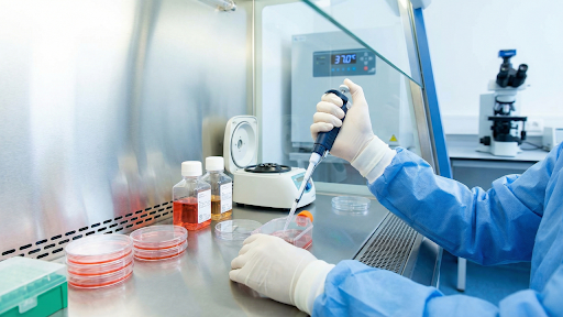
The modern biolab is marked by its characteristic of having spaces with no contamination. A row of incubators that keep millions of cells alive at 37°C.
Many of these cells are used in various ways, including to test cancer drugs, grow artificial skin, and produce life-saving vaccines. Microorganisms such as bacteria easily float in broth, but mammalian cells are “adherent.” They usually stick and sometimes get contaminated.
This leads to a problem for many researchers and a critical challenge that needs to be addressed: How do you move these cells when they run out of space?
This can be resolved by a technique called “passaging,” and with the help of biological tools such as chemical scissors. Let’s dissect the phenomenon in order.
The Process Of Holding Cells
The sticking as well as the loosening of cells are delicate processes. In case of sticking, the ECM (extracellular matrix) of the mammalian cell acts like an anchor, thus binds to the outer plastic of the flask with the help of adhesion proteins. Through the state of “confluency,” the divided cells cover almost the entire surface area of the flask. Researchers keep a keen eye and don’t let cells be in this state for a long time, as they could die due to contact inhibition and a lack of nutrients. To avoid and keep cells alive, they must be diluted and then moved to a fresh container.
Just scrapping them will lead to severe consequences, such as breaking of the cell’s outer membrane and ultimately killing them. Hence, the detachment process needs chemical scissors.
Leveraging Chemical Scissors
Now that we understand the importance of chemical scissors for cell detachment, a key molecular scissor is “Porcine Trypsinogen.” The nature of the molecular tool is “zymogen” – an inactive precursor produced in the pancreas of a pig.
The activated mature form of it is “Trypsin.” The nature of the mature form is a serine protease (a protein-digesting enzyme).
- Target: The Peptide bond of the carboxyl side of amino acids “Lysine” and “Arginine” is specifically targeted.
- Action: It is added to the culture flask because the enzyme cuts down the adhesion proteins that hold the cells to the plastic part of the flask.
- Result: It results in the retraction of the cells effectively. They eventually get detached, round up, and scientists collect them from the liquid area where they float.
Porcine-derived trypsin is the industry standard for this process because of its high efficacy and specificity compared to other sources. It effectively “frees” the cells without penetrating the membrane and destroying the internal machinery, provided the exposure time is strictly controlled.
Step-by-Step Workflow of “Passage”
A proper sterile technique, when met with precision, delivers a successful cell maintenance. Here is the step-by-step procedure:
01. Wash: Old nutrient media are required to be removed. It requires washing of cells with a saline solution such as PBS to remove all traces of serum. The objective is to be devoid of any traces of serum because it contains proteins that inhibit or restrict the working of trypsin.
02. Digestion: To achieve this, a solution of activated Porcine Trypsinogen (often combined with EDTA to help loosen cell-to-cell bonds) is added, and then the flask is placed back in the incubator for a limited period of time.
03. Tap/Handling: After washing and digestion, the researcher taps the flask. The handling must be done gently to avoid any kind of damage and contamination. The confirmation of flat cell conversion to smooth, floating spheres is observed under a microscope.
04. Removal: The addition of fresh serum-rich media is followed after confirmation of healthy cells. The inhibitors in the serum stop the trypsin from working, preventing it from digesting the cells themselves.
Tools and Quality Control
The phenomenon of passing is simple and straightforward. The tools and reagents involved must be of the highest quality because alteration in enzyme activity can lead to “over-trypsinization” and “under-trypsinization”.
In over-trypsinization, the cell membrane is damaged, while in under-trypsinization, the clumps of cells remain stuck, which leads to the uneven growth of cells. This not only ensures the success of passing but also the integrity of the experiment with reproducible results.
Companies like AAABio support this ecosystem by providing the validated antibodies, proteins, and ELISA kits that scientists rely on to benchmark their experiments and ensure the integrity of their biological models.
Final Thoughts
Maintaining the cell line and cell culture is one of the foundational pillars of life sciences. It opens doors for the analysis of various aspects of the human body.
From the study of molecular biology to genetic engineering and virology, the maintenance of cell lines has led to the discovery of many medical procedures and treatments. Hence, the maintenance of cell lines is not only important for the success of experiments but for the overall advancement of scientific research.